Tori M Saneda
Note: This chapter contains images of human skeletons. Readers who have concerns about viewing human remains should consult with their instructor.
Skeletal Anatomy
The study of bones is called osteology. It is necessary to have a basic understanding of skeletal anatomy since the data used in the study of primate and human evolution is based on fossilized bones. The information presented here focuses on the human skeleton.
Human beings have an endoskeleton, which means that the skeletal structure is on the inside of the body. The endoskeleton grows along with the rest of the body. Endoskeletons are lighter than exoskeletons, meaning the skeletal system is on the outside of the body. This lighter endoskeleton allowed for the evolution of large vertebrate animals. The endoskeleton is comprised of bone. Bone has three structural parts:
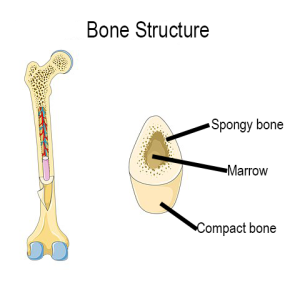
- Compact bone (periosteum): dense outer layer
- Spongy bone: light, softer inner layer
- Marrow: fills the inside core and makes red and white blood cells.
For the skeletal system to work properly it must have ligaments that connect the bone at the joints, muscles connected to the bones with tendons that allow for movement of the bone, and cartilage that fills the spaces between the bones, preventing them from scraping against one another.
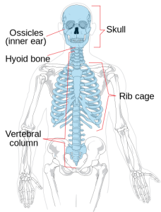
Human infants have 270 bones, several of which fuse together during growth so that adult humans have 206 bones. The skeleton is divided into two categories: the axial skeleton and the appendicular skeleton. The axial skeleton is comprised of the vertebral column, ribs, sternum, and skull.
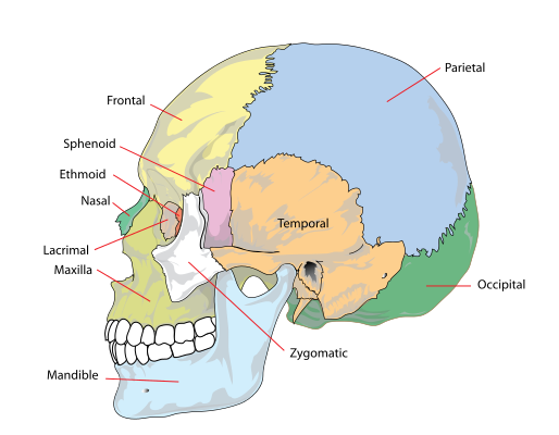
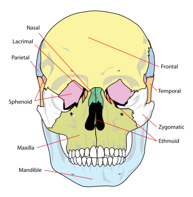
The human skull has a number of bones. These bones were separate at birth and then fuse together as an individual ages.
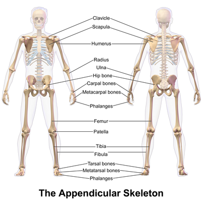
The appendicular skeleton is composed of the shoulder girdle, upper limbs, pelvic girdle, and the lower limbs.
Function of Bone
Bones function to:
- give structural support to the body
- protect vital organs
- provide an environment for the production of red blood cells
- store minerals like calcium
- aid in movement of the body
Bone Types
There are four types of bones based on shape.
- Long bones: long central shaft (diaphysis) and two knobby ends (epiphysis); e.g., femur
- Short bones: about as long as wide; e.g., patella
- Flat bones: characterized by being relatively thin and flat, e.g., sternum
- Irregular bones: odd-shaped bones; e.g., vertebrae
Bones can also be classified based on their origin and texture, but for the purposes of this class you need only be familiar with the types based on shape.
You should spend some time on the website eSkeletons in order to familiarize yourself with the look of the various bones listed above.
Types of Bone Cells
Osteoblast: forms new bone tissue
Osteoclast: absorbs and removes unwanted tissue
Osteocyte: maintains bone as living tissue
Hematopoietic: produces red blood cells
References
Carnagie J, Bruno LC, editors. 2001. Skeletal system. Detroit (MI): UXL. p. 538-542. (UXL Complete Life Science Resource; vol. 3).
Martin EL. 2014. Skeletal system. In: Lerner KL , Lerner BW, editors. The Gale Encyclopedia of Science, 5th edition, Vol. 7. Farmington Hills (MI): Gale. p. 3975-3981.
University of Chicago Medicine. c2015. Anatomy of the bone. The University of Chicago Medical Center [Internet] [cited 2015 Aug 2]. Available from: http://www.uchospitals.edu/online-library/content=P00109
Cite this page
APA Style: Saneda, T. M. 2022. Skeletal antaomy. In T. M. Saneda & M. Field, Biological Anthropology: a brief introduction. Cascadia College Pressbooks.
Chicago Style: Saneda, Tori M. 2022. “Skeletal Anatomy.” In Biological Anthropology: A Brief Introduction, 3rd. Bothell, WA: Cascadia College Pressbooks.
CSE Style: Saneda TM. 2010. Skeletal anatomy. In: Biological Anthropology: a brief introduction, 3rd ed. Bothell (WA): Cascadia College Pressbooks. [modified 2022; accessed 2022 Dec 5]. https://openwa.pressbooks.pub/anth205bioanth/chapter/skeletalanatomy.
