7.3 Prokaryotic Diversity
Learning Objectives
By the end of this section, you will be able to:
- Describe the evolutionary history of prokaryotes
- Describe the basic structure of a typical prokaryote
- Identify bacterial diseases that caused historically important plagues and epidemics
- Describe the uses of prokaryotes in food processing and bioremediation
Prokaryotes are present everywhere. They cover every imaginable surface where there is sufficient moisture, and they live on and inside of other living things. There are more prokaryotes inside and on the exterior of the human body than there are human cells in the body. Some prokaryotes thrive in environments that are inhospitable for most other living things. Prokaryotes recycle nutrients—essential substances (such as carbon and nitrogen)—and they drive the evolution of new ecosystems, some of which are natural while others are man-made. Prokaryotes have been on Earth since long before multicellular life appeared.
Prokaryotic Diversity
The advent of DNA sequencing provided immense insight into the relationships and origins of prokaryotes that were not possible using traditional methods of classification. A major insight identified two groups of prokaryotes that were found to be as different from each other as they were from eukaryotes. This recognition of prokaryotic diversity forced a new understanding of the classification of all life and brought us closer to understanding the fundamental relationships of all living things, including ourselves.
Early Life on Earth
When and where did life begin? What were the conditions on Earth when life began? Prokaryotes were the first forms of life on Earth, and they existed for billions of years before plants and animals appeared. Earth is about 4.54 billion years old. This estimate is based on evidence from the dating of meteorite material, since surface rocks on Earth are not as old as Earth itself. Most rocks available on Earth have undergone geological changes that make them younger than Earth itself. Some meteorites are made of the original material in the solar disk that formed the objects of the solar system, and they have not been altered by the processes that altered rocks on Earth. Thus, the age of meteorites is a good indicator of the age of the formation of Earth. The original estimate of 4.54 billion years was obtained by Clare Patterson in 1956. His meticulous work has since been corroborated by ages determined from other sources, all of which point to an Earth age of about 4.54 billion years.
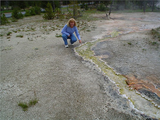
Early Earth had a very different atmosphere than it does today. Evidence indicates that during the first 2 billion years of Earth’s existence, the atmosphere was anoxic, meaning that there was no oxygen. Therefore, only those organisms that can grow without oxygen—anaerobic organisms—were able to live. Organisms that convert solar energy into chemical energy are called phototrophs. Phototrophic organisms that required an organic source of carbon appeared within one billion years of the formation of Earth. Then, cyanobacteria, also known as blue-green algae, evolved from these simple phototrophs one billion years later. Cyanobacteria are able to use carbon dioxide as a source of carbon. Cyanobacteria (Figure 13.2) began the oxygenation of the atmosphere. The increase in oxygen concentration allowed the evolution of other life forms.
Before the atmosphere became oxygenated, the planet was subjected to strong radiation; thus, the first organisms would have flourished where they were more protected, such as in ocean depths or beneath the surface of Earth. At this time, too, strong volcanic activity was common on Earth, so it is likely that these first organisms—the first prokaryotes—were adapted to very high temperatures. These are not the typical temperate environments in which most life flourishes today; thus, we can conclude that the first organisms that appeared on Earth likely were able to withstand harsh conditions.
Microbial mats may represent the earliest forms of life on Earth, and there is fossil evidence of their presence, starting about 3.5 billion years ago. A microbial mat is a large biofilm, a multi-layered sheet of prokaryotes (Figure 13.3a), including mostly bacteria, but also archaea. Microbial mats are a few centimeters thick, and they typically grow on moist surfaces. Their various types of prokaryotes carry out different metabolic pathways, and for this reason, they reflect various colors. Prokaryotes in a microbial mat are held together by a gummy-like substance that they secrete.
The first microbial mats likely obtained their energy from hydrothermal vents. A hydrothermal vent is a fissure in Earth’s surface that releases geothermally heated water. With the evolution of photosynthesis about 3 billion years ago, some prokaryotes in microbial mats came to use a more widely available energy source—sunlight—whereas others were still dependent on chemicals from hydrothermal vents for food.
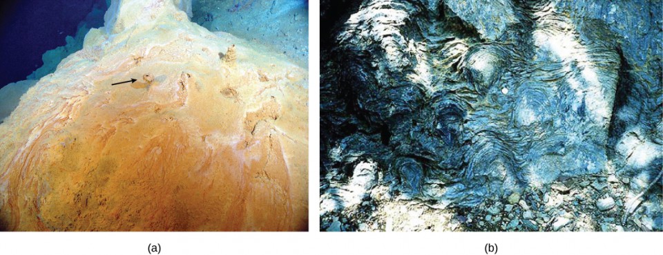
Fossilized microbial mats represent the earliest record of life on Earth. A stromatolite is a sedimentary structure formed when minerals are precipitated from water by prokaryotes in a microbial mat (Figure 13.3b). Stromatolites form layered rocks made of carbonate or silicate. Although most stromatolites are artifacts from the past, there are places on Earth where stromatolites are still forming. For example, living stromatolites have been found in the Anza-Borrego Desert State Park in San Diego County, California.
Some prokaryotes are able to thrive and grow under conditions that would kill a plant or animal. Bacteria and archaea that grow under extreme conditions are called extremophiles, meaning “lovers of extremes.” Extremophiles have been found in extreme environments of all kinds, including the depths of the oceans, hot springs, the Arctic and the Antarctic, very dry places, deep inside Earth, harsh chemical environments, and high radiation environments. Extremophiles give us a better understanding of prokaryotic diversity and open up the possibility of the discovery of new therapeutic drugs or industrial applications. They have also opened up the possibility of finding life in other places in the solar system, which have harsher environments than those typically found on Earth. Many of these extremophiles cannot survive in moderate environments.
Concept in Action

Watch a video showing the Director of the Planetary Science Division of NASA discussing the implications that the existence extremophiles on Earth have on the possibility of finding life on other planets in our solar system, such as Mars.
Biofilms
Until a couple of decades ago, microbiologists thought of prokaryotes as isolated entities living apart. This model, however, does not reflect the true ecology of prokaryotes, most of which prefer to live in communities where they can interact. A biofilm is a microbial community held together in a gummy-textured matrix, consisting primarily of polysaccharides secreted by the organisms, together with some proteins and nucleic acids. Biofilms grow attached to surfaces. Some of the best-studied biofilms are composed of prokaryotes, although fungal biofilms have also been described.
Biofilms are present almost everywhere. They cause the clogging of pipes and readily colonize surfaces in industrial settings. They have played roles in recent, large-scale outbreaks of bacterial contamination of food. Biofilms also colonize household surfaces, such as kitchen counters, cutting boards, sinks, and toilets.
Interactions among the organisms that populate a biofilm, together with their protective environment, make these communities more robust than are free-living, or planktonic, prokaryotes. Overall, biofilms are very difficult to destroy, because they are resistant to many of the common forms of sterilization.
Characteristics of Prokaryotes
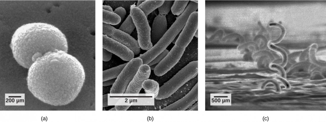
There are many differences between prokaryotic and eukaryotic cells. However, all cells have four common structures: a plasma membrane that functions as a barrier for the cell and separates the cell from its environment; cytoplasm, a jelly-like substance inside the cell; genetic material (DNA and RNA); and ribosomes, where protein synthesis takes place. Prokaryotes come in various shapes, but many fall into three categories: cocci (spherical), bacilli (rod-shaped), and spirilla (spiral-shaped) (Figure 13.5).
The Prokaryotic Cell
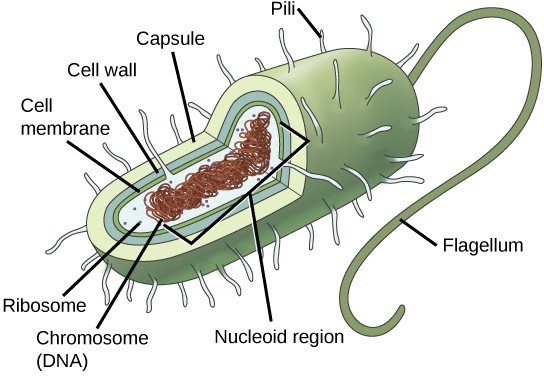
Recall that prokaryotes (Figure 13.5) are unicellular organisms that lack organelles surrounded by membranes. Therefore, they do not have a nucleus but instead have a single chromosome—a piece of circular DNA located in an area of the cell called the nucleoid. Most prokaryotes have a cell wall lying outside the plasma membrane. The composition of the cell wall differs significantly between the domains Bacteria and Archaea (and their cell walls also differ from the eukaryotic cell walls found in plants and fungi.) The cell wall functions as a protective layer and is responsible for the organism’s shape. Some other structures are present in some prokaryotic species, but not in others. For example, the capsule found in some species enables the organism to attach to surfaces and protects it from dehydration. Some species may also have flagella (singular, flagellum) used for locomotion, and pili (singular, pilus) used for attachment to surfaces and to other bacteria for conjugation. Plasmids, which consist of small, circular pieces of DNA outside of the main chromosome, are also present in many species of bacteria.
Both Bacteria and Archaea are types of prokaryotic cells. They differ in the lipid composition of their cell membranes and in the characteristics of their cell walls. Both types of prokaryotes have the same basic structures, but these are built from different chemical components that are evidence of an ancient separation of their lineages. The archaeal plasma membrane is chemically different from the bacterial membrane; some archaeal membranes are lipid monolayers instead of phosopholipid bilayers.
The Cell Wall
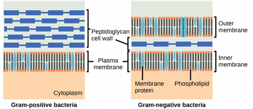
The cell wall is a protective layer that surrounds some prokaryotic cells and gives them shape and rigidity. It is located outside the cell membrane and prevents osmotic lysis (bursting caused by increasing volume). The chemical compositions of the cell walls vary between Archaea and Bacteria, as well as between bacterial species. Bacterial cell walls contain peptidoglycan, composed of polysaccharide chains cross-linked to peptides. Bacteria are divided into two major groups: Gram-positive and Gram-negative, based on their reaction to a procedure called Gram staining. The different bacterial responses to the staining procedure are caused by cell wall structure. Gram-positive organisms have a thick wall consisting of many layers of peptidoglycan. Gram-negative bacteria have a thinner cell wall composed of a few layers of peptidoglycan and additional structures, surrounded by an outer membrane (Figure 13.6).
Visual Connection
Which of the following statements is true?
- Gram-positive bacteria have a single cell wall formed from peptidoglycan.
- Gram-positive bacteria have an outer membrane.
- The cell wall of Gram-negative bacteria is thick, and the cell wall of Gram-positive bacteria is thin.
- Gram-negative bacteria have a cell wall made of peptidoglycan, while Gram-positive bacteria have a cell wall made of phospholipids.
(Answer: 1)
Archaeal cell walls do not contain peptidoglycan. There are four different types of archaeal cell walls.
Reproduction
Reproduction in prokaryotes is primarily asexual and takes place by binary fission. Recall that the DNA of a prokaryote exists usually as a single, circular chromosome. The chromosome loop is replicated, and the two resulting copies attached to the plasma membrane move apart as the cell grows in a process called binary fission. The prokaryote, now enlarged, is pinched inward at its equator, and the two resulting cells, which are clones, separate. Binary fission does not provide an opportunity for genetic recombination, but prokaryotes can alter their genetic makeup in three ways.
In a process called transformation, the cell takes in DNA found in its environment that is shed by other prokaryotes, alive or dead. A pathogen is an organism that causes a disease. If a nonpathogenic bacterium takes up DNA from a pathogen and incorporates the new DNA in its own chromosome, it too may become pathogenic. In transduction, bacteriophages, the viruses that infect bacteria, move DNA from one bacterium to another. Archaea have a different set of viruses that infect them and translocate genetic material from one individual to another. During conjugation, DNA is transferred from one prokaryote to another by means of a pilus that brings the organisms into contact with one another. The DNA transferred is usually a plasmid, but parts of the chromosome can also be moved.
Cycles of binary fission can be very rapid, on the order of minutes for some species. This short generation time coupled with mechanisms of genetic recombination result in the rapid evolution of prokaryotes, allowing them to respond to environmental changes (such as the introduction of an antibiotic) very quickly.
How Prokaryotes Obtain Energy and Carbon
Prokaryotes are metabolically diverse organisms. Prokaryotes fill many niches on Earth, including being involved in nutrient cycles such as the nitrogen and carbon cycles, decomposing dead organisms, and growing and multiplying inside living organisms, including humans. Different prokaryotes can use different sources of energy to assemble macromolecules from smaller molecules. Phototrophs obtain their energy from sunlight. Chemotrophs obtain their energy from chemical compounds.
Beneficial Prokaryotes
Not all prokaryotes are pathogenic. On the contrary, pathogens represent only a very small percentage of the diversity of the microbial world. In fact, our life and all life on this planet would not be possible without prokaryotes.
Prokaryotes, and Food and Beverages
According to the United Nations Convention on Biological Diversity, biotechnology is “any technological application that uses biological systems, living organisms, or derivatives thereof, to make or modify products or processes for specific use.”4 The concept of “specific use” involves some sort of commercial application. Genetic engineering, artificial selection, antibiotic production, and cell culture are current topics of study in biotechnology. However, humans have used prokaryotes to create products before the term biotechnology was even coined. And some of the goods and services are as simple as cheese, yogurt, sour cream, vinegar, cured sausage, sauerkraut, and fermented seafood that contains both bacteria and archaea (Figure 13.9).
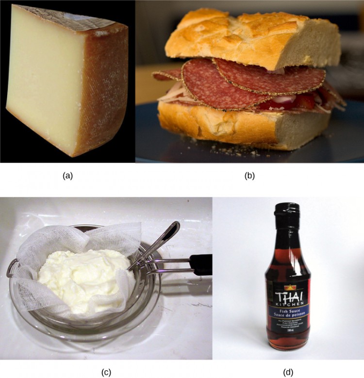
Cheese production began around 4,000 years ago when humans started to breed animals and process their milk. Evidence suggests that cultured milk products, like yogurt, have existed for at least 4,000 years.
Using Prokaryotes to Clean up Our Planet: Bioremediation
Microbial bioremediation is the use of prokaryotes (or microbial metabolism) to remove pollutants. Bioremediation has been used to remove agricultural chemicals (pesticides and fertilizers) that leach from soil into groundwater. Certain toxic metals, such as selenium and arsenic compounds, can also be removed from water by bioremediation. The reduction of SeO42- to SeO32-and to Se0 (metallic selenium) is a method used to remove selenium ions from water. Mercury is an example of a toxic metal that can be removed from an environment by bioremediation. Mercury is an active ingredient of some pesticides; it is used in industry and is also a byproduct of certain industries, such as battery production. Mercury is usually present in very low concentrations in natural environments but it is highly toxic because it accumulates in living tissues. Several species of bacteria can carry out the biotransformation of toxic mercury into nontoxic forms. These bacteria, such as Pseudomonas aeruginosa, can convert Hg2+ to Hg0, which is nontoxic to humans.
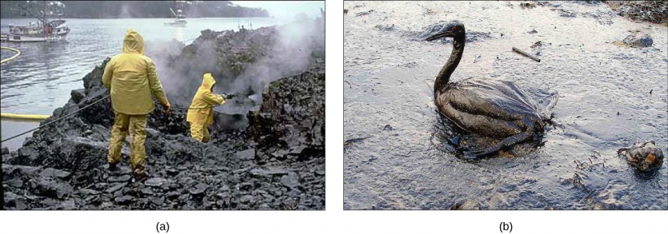
Probably one of the most useful and interesting examples of the use of prokaryotes for bioremediation purposes is the cleanup of oil spills. The importance of prokaryotes to petroleum bioremediation has been demonstrated in several oil spills in recent years, such as the Exxon Valdez spill in Alaska (1989) (Figure 13.10), the Prestige oil spill in Spain (2002), the spill into the Mediterranean from a Lebanon power plant (2006,) and more recently, the BP oil spill in the Gulf of Mexico (2010). To clean up these spills, bioremediation is promoted by adding inorganic nutrients that help bacteria already present in the environment to grow. Hydrocarbon-degrading bacteria feed on the hydrocarbons in the oil droplet, breaking them into inorganic compounds. Some species, such as Alcanivorax borkumensis, produce surfactants that solubilize the oil, while other bacteria degrade the oil into carbon dioxide. In the case of oil spills in the ocean, ongoing, natural bioremediation tends to occur, inasmuch as there are oil-consuming bacteria in the ocean prior to the spill. Under ideal conditions, it has been reported that up to 80 percent of the nonvolatile components in oil can be degraded within 1 year of the spill. Other oil fractions containing aromatic and highly branched hydrocarbon chains are more difficult to remove and remain in the environment for longer periods of time. Researchers have genetically engineered other bacteria to consume petroleum products; indeed, the first patent application for a bioremediation application in the U.S. was for a genetically modified oil-eating bacterium.
Prokaryotes in and on the Body
Humans are no exception when it comes to forming symbiotic relationships with prokaryotes. We are accustomed to thinking of ourselves as single organisms, but in reality, we are walking ecosystems. There are 10 to 100 times as many bacterial and archaeal cells inhabiting our bodies as we have cells in our bodies. Some of these are in mutually beneficial relationships with us, in which both the human host and the bacterium benefit, while some of the relationships are classified as commensalism, a type of relationship in which the bacterium benefits and the human host is neither benefited nor harmed.
Human gut flora lives in the large intestine and consists of hundreds of species of bacteria and archaea, with different individuals containing different species mixes. The term “flora,” which is usually associated with plants, is traditionally used in this context because bacteria were once classified as plants. The primary functions of these prokaryotes for humans appear to be metabolism of food molecules that we cannot break down, assistance with the absorption of ions by the colon, synthesis of vitamin K, training of the infant immune system, maintenance of the adult immune system, maintenance of the epithelium of the large intestine, and formation of a protective barrier against pathogens.
The surface of the skin is also coated with prokaryotes. The different surfaces of the skin, such as the underarms, the head, and the hands, provide different habitats for different communities of prokaryotes. Unlike with gut flora, the possible beneficial roles of skin flora have not been well studied. However, the few studies conducted so far have identified bacteria that produce antimicrobial compounds as probably responsible for preventing infections by pathogenic bacteria.
Researchers are actively studying the relationships between various diseases and alterations to the composition of human microbial flora. Some of this work is being carried out by the Human Microbiome Project, funded in the United States by the National Institutes of Health.
Section Summary
Prokaryotes existed for billions of years before plants and animals appeared. Microbial mats are thought to represent the earliest forms of life on Earth, and there is fossil evidence, called stromatolites, of their presence about 3.5 billion years ago. During the first 2 billion years, the atmosphere was anoxic and only anaerobic organisms were able to live. Cyanobacteria began the oxygenation of the atmosphere. The increase in oxygen concentration allowed the evolution of other life forms.
Prokaryotes (domains Archaea and Bacteria) are single-celled organisms lacking a nucleus. They have a single piece of circular DNA in the nucleoid area of the cell. Most prokaryotes have cell wall outside the plasma membrane. Bacteria and Archaea differ in the compositions of their cell membranes and the characteristics of their cell walls.
Bacterial cell walls contain peptidoglycan. Archaean cell walls do not have peptidoglycan. Bacteria can be divided into two major groups: Gram-positive and Gram-negative. Gram-positive organisms have a thick cell wall. Gram-negative organisms have a thin cell wall and an outer membrane. Prokaryotes use diverse sources of energy to assemble macromolecules from smaller molecules. Phototrophs obtain their energy from sunlight, whereas chemotrophs obtain it from chemical compounds.
Prokaryotes are used in human food products. Microbial bioremediation is the use of microbial metabolism to remove pollutants. The human body contains a huge community of prokaryotes, many of which provide beneficial services such as the development and maintenance of the immune system, nutrition, and protection from pathogens.
Glossary
- anaerobic: refers to organisms that grow without oxygen
- anoxic: without oxygen
- biofilm: a microbial community that is held together by a gummy-textured matrix
- bioremediation: the use of microbial metabolism to remove pollutants
- capsule: an external structure that enables a prokaryote to attach to surfaces and protects it from dehydration
- commensalism: a symbiotic relationship in which one member benefits while the other member is not affected
- conjugation: the process by which prokaryotes move DNA from one individual to another using a pilus
- cyanobacteria: bacteria that evolved from early phototrophs and oxygenated the atmosphere; also known as blue-green algae
- extremophile: an organism that grows under extreme or harsh conditions
- Gram-negative: describes a bacterium whose cell wall contains little peptidoglycan but has an outer membrane
- Gram-positive: describes a bacterium that contains mainly peptidoglycan in its cell walls
- hydrothermal vent: a fissure in Earth’s surface that releases geothermally heated water
- microbial mat: a multi-layered sheet of prokaryotes that may include bacteria and archaea
- pathogen: an organism, or infectious agent, that causes a disease
- peptidoglycan: a material composed of polysaccharide chains cross-linked to unusual peptides
- phototroph: an organism that uses energy from sunlight
- pseudopeptidoglycan: a component of some cell walls of Archaea
- stromatolite: a layered sedimentary structure formed by precipitation of minerals by prokaryotes in microbial mats
- transduction: the process by which a bacteriophage moves DNA from one prokaryote to another
- transformation: a mechanism of genetic change in prokaryotes in which DNA present in the environment is taken into the cell and incorporated into the genome
Footnotes
- 1 Papagrigorakis M. J., Synodinos P. N., Yapijakis C, “Ancient typhoid epidemic reveals possible ancestral strain of Salmonella enterica serovar Typhi, Infect Genet Evol 7 (2007): 126-7.
- 2 Naimi, T. S., LeDell, K. H., Como-Sabetti, K., et al., “Comparison of community- and health care-associated methicillin-resistant Staphylococcus aureus infection,” JAMA 290 (2003): 2976-2984, doi: 10.1001/jama.290.22.2976.
- 3 http://www.cdc.gov/ecoli/2006/september, Centers for Disease Control and Prevention, “Multi-state outbreak of E. coli O157:H7 infections from spinach,” September-October (2006).
- 4 http://www.cbd.int/convention/articles/?a=cbd-02http://www.cbd.int/convention/articles/?a=cbd-02, United Nations Convention on Biological Diversity, “Article 2: Use of Terms.”
Media Attributions
- Figure 13.2
- Figure 13.3
- 13.1QR
- Figure 13.5
- Figure 13.6
- Figure 13.6
- Figure 13.9
- Figure 13.10
