5 Lab 4. Prokaryotic and Eukaryotic Cells
LAB 4—Prokaryotic & Eukaryotic Cells
OBJECTIVES
- Explore cell structure and morphology in prokaryotes and eukaryotes.
- Gain more experience using the microscope.
- Define the terms used in this lab: prokaryote, eukaryote, cell, cell membrane, cell wall, nucleus, mitochondrion.
- Give two general characteristics of prokaryotic cells.
- Identify the subcellular structures of a typical plant cell and review its function(s).
- Identify the subcellular structures of a typical animal cell and review its function(s).
- Compare and contrast prokaryotic and eukaryotic cells (i.e., be able to describe similarities and differences).
- Compare and contrast animal and plant cells.
PRELAB
- Read lab 4 and be ready to begin the lab exercise.
- Understand the primary differences between prokaryotic and eukaryotic cells.
- In your lab notebook diagram what you will do in Lab 4. This can take many different forms: flow chart, diagram, series of cartoons, list, outline, etc. It is important that you know what you are going to do and plan your time BEFORE you come to lab.
- The Biology Project at the University of Arizona has an excellent website that may be useful to you at different times during the quarter. The following page has a good, simple review of light microscopy, electron microscopy and size of cells: Studying Cells Tutorial (arizona.edu)
INTRODUCTION
Understanding the nature of cell structure and function is important to understanding organisms. All organisms are composed of cells, whether they exist as single cells, colonies of cells, or in multicellular form. Cells are usually very small, and for this reason, a thorough understanding of subcellular structure and function has been possible only through advances in electron microscopy and molecular biology.
There are two general types of cells: prokaryotic and eukaryotic. These two words have their root in the Greek word karyon (nut), which refers to a cell’s nucleus. The prefix pro- means “before”. Thus prokaryotic means “before having a nucleus.” Prokaryotic cells do not have a nucleus and their genetic material (DNA) is in a nuclear area within the cell. Archaea and bacteria, including cyanobacteria (formerly known as blue-green algae) are prokaryotes. All other organisms are eukaryotes. The prefix “eu”- means “true.” The cells of eukaryotes have true, double-membrane nuclei containing their genetic material (DNA).
Prokaryotic and eukaryotic cells differ in several ways. Eukaryotic cells are generally larger and contain membranous organelles with specialized functions. Prokaryotic cells lack membrane-bounded organelles; their cell functions are carried out in the cytoplasm.
In this laboratory, you will investigate some of the structural properties of prokaryotic and eukaryotic cells. We are going to focus on the primary differences between eukaryotes and prokaryotes. In this lab, you will examine cells from two domains: Bacteria and Eukarya and two Eukaryotic kingdoms: Plantae and Animalia. You should work in groups of two.
PROCEDURE
Part A: Prokaryotic cells
Present-day bacteria are found everywhere: in soil, in water, in ice, in boiling hot pools of water, even kilometers underground! Many depend on food in the form of organic compounds (heterotrophic), some make their own food with light (photosynthetic), and some are capable of using inorganic substances as their source of energy – they essentially “eat rock” (lithotrophic). Bacteria are extremely small (approximately 1 to 2 µm in diameter). To view them with the light microscope, one must use at least the high dry objective (400x). Even then, not much more than their basic shapes are visible. To learn more with the light microscope, one can use special staining techniques. With the aid of the electron microscope, however, one can study these prokaryotic cells in much greater detail.
Observing Bacteria
In this lab, we will observe cyanobacteria, a type of bacteria commonly found in soils and waters in the environment.
- Make a wet mount of the cyanobacteria species (Anabaena spp.) in the lab and observe first under 40x, then 100x, and then 400x (using the high-dry objective). Using the scanning (4x objective, 40x total magnification), they will appear as very small green lines. You may need assistance locating these. Then you will center the image and focus
before moving to higher magnification. These species form thread-like colonies. Refer to the picture below of Anabaena.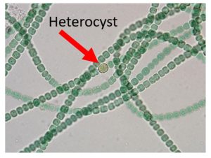
- At 400x, you should be able to see two different types of cells making up a colony.
- Draw a representative colony (a strand of cells) showing these two types. Include the name of the organism and the total magnification.
- Find and draw a strand with at least two heterocysts. Count and record the number of “vegetative” cells between two heterocysts.
- In a sentence or two, compare and contrast “vegetative” cells and heterocysts (with respect to size, shape, color, number, etc.).
Part B: Eukaryotic cells
All eukaryotic organisms are composed of cells, whether they exist as single cells, colonies of cells, or in multicellular form. Your body is composed of 50 to 100 trillion cells, most of which are very small, with specialized structures that allow for a diversity of functions. Plants, animals, fungi and protists (including many single-celled organisms) are all eukaryotes. All eukaryotic cells have their genetic material enclosed by a double membrane called the nuclear envelope. In addition, a variety of subcellular structures bounded by a membrane, called membrane-bounded organelles, are present. These include plastids, mitochondria, lysosomes, microbodies, and Golgi complexes. Internal membrane systems, or endoplasmic reticula, divide the cell into specialized compartments. Non-membrane-bounded organelles such as ribosomes, centrioles, microtubules, and microfilaments, are also present in eukaryotic cells.
The following structures can be seen in some, but not all, eukaryotic cells. Review and be able to describe the structure and function of the following cell structures. Refer to the plant and animal cell models and diagrams in the lab room, the photo atlases, and your textbook. You do NOT need to list these in your lab notebook. However, you may need to label some of these these structures in some of your drawings (where appropriate).
plasma membrane (or cell membrane), cell wall, chloroplasts, leucoplasts (also called amyloplasts), vacuoles, including the central vacuole in plant cells, cytoplasm, flagella (or flagellum, singular), cilia, nucleus, nucleolus
NOTE: You will not be able to see most of the structures listed above in your observations of actual cells using the light microscope – they are too small or may not be visible unless they are stained. Instead refer to posters, images and your textbook.
Part 1: Examining Plant Cells
Plants are multicellular organisms composed of eukaryotic cells. Plant cells contain a large central vacuole surrounded by a membrane stores water, pigments and wastes. Within the cytoplasm are different types of plastids. These may include the green-colored chloroplast, responsible for photosynthesis, and amyloplasts (or leukopasts) that store starch. A cell wall surrounds the cell membrane and is composed of cellulose; this surrounds the plant cell.
Leaf of an aquatic green plant. (See image below for photograph of Elodea.) Elodea is a genus name, this name (and other genus names) must be Capitalized and underlined in your lab notebook. You will choose another aquatic green plant leaf to observe. There will be three or four choices available during your lab section, including another species in the genus Elodea, subwassertang, an aquarium plant belonging to a species in the Genus Lomariopsis and an aquarium grass.
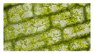
A. Thin green leaf of an aquatic green plant.
- Prepare a wet-mount slide of an Elodea or other green leaf.
- View the leaf cells using the scanning objective. Find the leaf, focus and center the image, then move to the low power (10x) objective, and then high power (40x) objective. Observe the thick cell wall, thinner cell membrane (this structure may not be visible between the cell wall and the cytoplasm), cytoplasm, and chloroplasts. A large central vacuole should be apparent. These structures characterize a generalized plant cell. In some leaf cells you may be able to see the nucleus.
- Sketch a representative leaf cell as observed under high power and label its parts. Include the name of the organism and the TOTAL magnification in your drawing. Answer the following questions in your lab notebook:
- Note whether the chloroplasts appear to move. If so, describe their movement.
- Are all of the leaf cells in your field of view the same size and shape?
B. Potato
- Use a razor blade or peeler to slice a piece of tissue, as thin as possible, from a potato. Be careful not to cut your fingers. Prepare a wet-mount slide, using a drop of water and a coverslip.
- Study the slide at low power (10x objective) and then at high power (40x objective). Add a drop of Lugol’s solution (iodine or I2KI) to the side of the coverslip and touch a piece of Kimwipe to the other side of the coverslip to draw the stain solution under the coverslip. The iodine will stain starch a very deep blue/purple/black. Observe the cells as the iodine solution makes contact with them. Answer the following questions in your lab notebook:
- What do your observations tell you about structure and possible function of these cells? Plants store excess energy as molecules.
- Do you see any chloroplasts? If so, why would the potato cells have them? If not, why would the potato cells lack them?
- Do you see small oval-shaped structures (leucoplasts) within the cells? What color are they? What does their color indicate?
- Observe the cell walls of these cells? Compare and contrast the potato and leaf cell with respect to size, shape, structures and organelles.
- Sketch a representative stained potato cell as observed under high power and label the cell wall, leucoplasts and starch. Again, be sure to include the name of the organism and the total magnification. How large are these cells?
In your lab notebook, make a table describing the three (3) similarities and three (3) differences between green plant cells and potato cells. You may complete this table and paste it into your lab notebook.
Table 1. Similarities and differences between green plant cells and potato cells.
| Similarities | Differences |
|
|
|
|
|
|
|
|
Part 2: Examining Animal Cells
Animal cells can be studied using the light microscope, but most of the cellular organelles within the cytoplasm are not visible without the use of special staining techniques. After staining with methylene blue, the nucleus and nucleolus, where ribosomes are manufactured, are usually apparent in most cells.
Cheek cells
You will examine cheek cells obtained from your mouth (human epithelial cells). Because cheek cells are considered biohazardous material, please follow the clean-up instructions carefully!
Prepare a “wet mount” slide with of your cheek cells.
- Obtain a clean glass slide (you may want to clean it again yourself with a Kimwipe). Be careful to hold the slide by the edges to avoid smudging it.
- Place one (1) drop each of water & methylene blue in the center of the slide.
- Gently rub the inside of your cheek with the blunt end of a toothpick.
- Stir the cheek material (you may not be able to see any) into the methylene blue in the center of the slide. If the stain appears very dark, add a drop of water.
- Apply a cover slip; touch one edge of the cover slip to the edge of the drop of liquid. Hold the coverslip while the liquid runs along the entire edge. Then gently lower the coverslip over the specimen.
- Dispose of toothpick in the beaker of bleach provided.
- Place the cheek cell slide on the microscope and examine it under the scanning objective. Focus the cells, adjust the light intensity, and especially adjust the contrast using the iris diaphragm lever.
- Examine your cheek cells. NOTE: These cells are flat with irregular shapes or contours. Some of the thin, flat edges of the cells may be folded in your preparation. Many of the cells will probably be clumped together, which makes observing individual cells difficult. Bacteria may also be present as small, dot-like or stick-like structures covering the cells.
- Find an area of the slide where the cells are separate from each other. Select cells for closer examination that have centrally-located, darkly-stained nuclei.
- Observe these cells using the low power objective. Move the slide so that an individual cell is in the center of the field of view. Then move the high power objective into place and bring the cell into focus using the fine-focus knob. Draw a single, isolated squamous cheek cell. Find and label the following structures: plasma (cell) membrane, cytoplasm, and nucleus.
- Dispose of the contaminated slides with the cover slips in the beaker of bleach solution.
Complete the following table in your lab notebook and note three (3) similarities and three (3) differences between the plant cells and the animal cells you have observed. You may complete this table and paste it into your lab notebook.
Table 2. Similarities and differences between plant cells and animal cells.
| Similarities | Differences |
|
|
|
|
|
|
|
|
CONCLUSION
In a short paragraph, discuss the similarities and differences you observed between prokaryotic and eukaryotic cells.
Clean up:
- Put away the microscope as instructed in Lab 1.
- Replace all other materials (slides, posters, etc.) to their proper location.
- The used cheek slides, cover slips, and other materials that touched the cheek cells should be put in the appropriate containers for sterilization.
- The other slides used for your other (plant) wet mount preparations should be washed, dried and returned to the slide boxes. Dispose of used cover slips in the broken glass box.
- Remove any debris from the sink(s) and put in the garbage.
- Clean and dry your lab bench (using paper towels).
Thank you for leaving the lab neat and clean!
