Medical Specialists
Cardiology (kăr-dē-ŏl′ō-jē) is the study of the heart. Specialty roles related to cardiology include cardiologists, cardiovascular surgeons, electrophysiologists, cardiovascular technologists and technicians, cardiovascular perfusionists, cardiac care nurses, cardiology nurse practitioners, and cardiology physician assistants.
Cardiologists
A cardiologist (kar-dē-OL-ŏ-jĭst) is a physician who specializes in the diagnosis and treatment of heart disease. Cardiovascular surgeons surgically treat heart disorders. An electrophysiologist is a physician who specializes in the diagnosis and treatment of issues related to the heart’s electrical conduction system.
Read more information about cardiologists on the American Medical Association’s Cardiovascular Disease Specialty web page.
Vascular Surgeon
Vascular surgery includes repair and replacement of diseased or damaged blood vessels, removal of plaque from vessels, and insertion of venous catheters, and traditional surgery.[1]
Vascular Sonographer
Vascular sonographers use ultrasound machines to produce images of patients’ veins and arteries using high-frequency sound waves. Physicians use these images that show the movement of blood through vessels to diagnose and treat various conditions.[2]
Cardiovascular Technologists and Technicians
Cardiovascular technologists and technicians perform cardiovascular diagnostic tests and procedures such as electrocardiography, stress testing, Holter monitor testing, ambulatory blood pressure (BP) testing, and pacemaker monitoring.
Read more about cardiovascular technologist and technician positions on the U.S. Bureau of Labor Statistics 29-2031 Cardiovascular Technologists and Technicians page.
Cardiovascular Perfusionist
Cardiovascular perfusionists operate circulation equipment that artificially support or temporarily replace a patient’s circulatory or respiratory function. An example of such equipment is a heart-lung machine used during some types of coronary artery bypass surgery.
Visit the Mayo Clinic: Cardiovascular Perfusionist web page for more information.
Cardiac Care Nurses
Cardiac care nurses provide care for patients with a variety of heart diseases or conditions in a cardiac care unit (CCU) in hospitals. They administer heart medications, help patients recover from heart surgery, or perform emergency care like assisting in defibrillation.
Cardiology Nurse Practitioners
Cardiology nurse practitioners (NPs) are registered nurses with advanced education and clinical experience to provide care for patients with chronic and acute cardiac diseases. Many cardiology NPs work in private practices, in-patient hospitals, outpatient clinic settings, or multiple practice settings where they assess the health status of patients, prescribe pharmacological and nonpharmacological treatments, and collaborate with an interdisciplinary team.
Read more information on “A Day in the Life of a Cardiology Nurse Practitioner (NP)” web page by the American Association of Nurse Practitioners.
Cardiology Physician Assistants
Cardiology physician assistants (PAs) work in collaboration with cardiologists and cardiovascular surgeons. They provide a broad range of medical care to patients with varied clinical duties depending on the subspecialty and setting. PAs take medical histories, perform physical examinations, order and interpret laboratory and diagnostic tests, diagnose illness, develop and manage treatment plans for their patients, prescribe medications, perform procedures, and assist in surgery.
Read more information about cardiology PAs on the “PAs in Cardiology” PDF from the American Academy of Physician Assistants website.
Radiology Technologist
A radiology technologist (rā-dē-ŎL-ŏ-jē tĕk-nŎL-ŏ-jĭst), commonly called an X-ray tech, is a health care professional who is specially trained to perform medical imaging like X-rays, CT scans, MRIs, and PET scans. To become a radiology technologist or MRI technologist, a person completes an associate degree and passes a certification exam.
Read more information about the occupational outlook for radiology technologists on the U.S. Bureau of Labor Statistics webpage.
Diagnostic Testing
Cardiac Catheterization and Angiogram
Cardiac catheterization (kär′dē-ăk kăth-ĕ-tĕr-ĭ-ZĀ-shŏn) is a valuable diagnostic procedure in the evaluation and management of cardiovascular disorders. Cardiac catheterization, performed in association with a coronary angiogram, is a diagnostic procedure used to visualize the coronary arteries, heart chambers, and great vessels. It involves the insertion of a thin, flexible tube (catheter) into the blood vessels, usually through the femoral or radial artery, to access the inside of the heart and its surrounding structures.
During a coronary angiogram (KOR-ŏ-nā-rē AN-jee-ō-gram), dye is injected into the blood vessels of the heart, and then a series of X-ray images (angiograms) are taken to view the coronary arteries. If a blockage is discovered, an angioplasty can be performed. Read more about angioplasty under the “Procedures” subsection later in this section.
See Figure 9.21[3] for an image of a cardiac catheterization procedure showing the image of the patient’s coronary arteries and heart chambers displayed on the monitor during the angiogram.

Cardiac Stress Test
A cardiac stress test (kär′-dē-ăk strĕs tĕst), also known as an exercise stress test or treadmill test, is a diagnostic procedure used to evaluate the performance and function of the heart during physical activity. It helps identify potential heart problems, such as coronary artery disease (CAD), arrhythmias, and heart valve issues. During a cardiac stress test procedure, a patient is asked to walk on a treadmill or pedal a stationary bicycle to evaluate the heart’s function during strenuous activity. In patient circumstances where walking on a treadmill is not appropriate, medication is administered intravenously to mimic the stress on the heart induced by exercise, referred to as a chemical stress test. See Figure 9.22[4] for an image of a cardiac stress test.

Electrocardiogram
An electrocardiogram (ē-lĕk-trō-KĂR-dē-ō-grăm) (EKG or ECG) is a diagnostic test that records the electrical activity of the heart. It is performed to diagnose a myocardial infarction (MI) or cardiac rhythm abnormalities. See Figure 9.23[5] for an image of a patient undergoing an ECG.

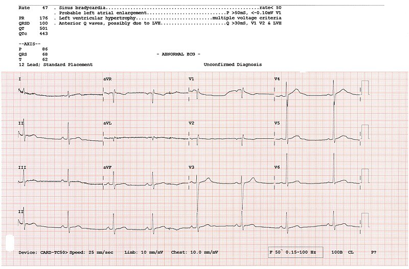
During an ECG, surface electrodes are placed on specific anatomical sites on the body to record electrical activity. An example of a 12-lead ECG is presented in Figure 9.24.[6]

Echocardiogram
An echocardiogram (ek-ō-KAR-dē-ō-gram) (ECHO) is a noninvasive diagnostic test that uses sound waves (ultrasound) to create real-time images of the heart’s structure and function. A trained sonographer or cardiologist applies a gel and a transducer to the patient’s chest. The transducer is moved to different areas of the chest to obtain various views of the heart, including the heart chambers, valves, walls, and blood flow patterns. Doppler ultrasound is also used during echocardiography to determine the speed and direction of blood flow through the valves and chambers of the heart. An echocardiogram is often used to diagnose heart failure and valve disorders impacting the flow of blood through and out of the heart.[7]
A transesophageal echocardiogram (trăn-sē-sŏf-ă-JĒ-ăl ek-ō-KAR-dē-ō-gram) (TEE) uses an ultrasound probe placed in the patient’s esophagus to provide a more direct view of heart structures.
Holter Monitor
A Holter monitor (HOL-tĕr MON-ĭ-tŏr) is a portable device that continuously records a patient’s heart rhythm and electrical activity over an extended period, typically 24 to 48 hours. See Figure 9.25[8] for an illustration of a Holter monitor. Its primary purpose is to detect and document irregularities in the heart’s electrical patterns, such as arrhythmias, which may not be captured during brief in-office ECGs. Additionally, patients may activate a trigger on a Holter device to denote the occurrence of cardiac symptoms, such as increased shortness of breath, fatigue, or dizziness. A cardiologist reviews ECG tracings to identify arrhythmias.

SPECT Scan
A single-photon emission computerized tomography scan (sĭng′-gŭl fō′-tŏn ē-mĭsh′-ŭn kŏm-pyū′-tə-rīzd tŏ-mŏg′-rə-fē skăn) (SPECT) analyzes the function of the body’s organs, tissues, and bones. For example, a SPECT scan can show how well blood is flowing in the coronary arteries or arteries in the brain. It can also show how completely the heart chambers empty during contractions. A SPECT scan is a type of nuclear imaging test, which means it uses a radioactive substance and a special camera to create 3D pictures. A radioactive substance is administered via intravenous (ĭn-tră-VĒ-nŭs) (IV) infusion (i.e., into a vein). The SPECT machine is a large circular device containing a camera that detects the radioactive tracer the body absorbs. During the scan, the patient lies on a table while the SPECT machine rotates around them and takes images of their internal organs and other structures. The pictures are sent to a computer that uses the information to create 3D images of the body.[9]
Procedures
Cardiac Ablation
Cardiac ablation (kär′dē-ăk ă-BLĀ-shŏn) uses heat or cold energy to create tiny scars in the heart to block irregular electrical signals and restore a typical heartbeat. This procedure is used to correct various types of arrhythmias. Cardiac ablation is most often done using thin, flexible tubes called catheters inserted through veins or arteries but can also be performed during cardiac surgery.[10]
Angioplasty
During a cardiac catheterization, if a blockage is found, an angioplasty (AN-jee-ō-plas-tē) may be performed, also referred to as a percutaneous transluminal coronary angioplasty (pĕr-kyū-TĀ-nē-ŭs trăns-LOO-mĭ-năl KOR-ŏ-nā-rē AN-jee-ō-plas-tē) (PTCA). During an angioplasty, a catheter with a balloon at the tip is inserted into the narrowed coronary artery. The balloon is inflated to widen the artery and improve blood flow. A small mesh device, known as a stent, is often inserted to help keep the coronary artery open after the procedure is completed. See Figure 9.26[11] for an illustration of an angioplasty with stent placement.

Defibrillators
Defibrillators can lower the risk of sudden death among people who have a known arrhythmia or a high risk of a life-threatening arrhythmia from causes such as genetics diseases, heart failure, or a prior cardiac arrest.
Defibrillators have different uses, depending on the type.
- Automated external defibrillators (AEDs) are found in many public spaces. They can save the lives of people who are in cardiac arrest. The unit tells the operator what to do. Even untrained people can use an AED in an emergency. Healthcare providers sometimes recommend keeping an AED at home if someone has a high risk for cardiac arrest.
- Implanted cardioverter defibrillators (ICDs) are small devices surgically placed in the chest. They are preprogramed to automatically detect cardiac arrest or a life-threatening arrhythmia. They then send a high-energy electric charge to stop the arrhythmia or restart the heart after cardiac arrest. Some ICDs also act as pacemakers by giving low-energy electrical pulses to help the heart beat at a normal rhythm. Others can send pulses of electricity to sync the rhythm of the heart’s lower chambers.
Coronary Artery Bypass Surgery
Coronary artery bypass (KOR-ŏ-nā-rē AR-tĕr-ē bī-păs) surgery, also called coronary artery bypass graft (KOR-ŏ-nā-rē AR-tĕr-ē bī-păs graft) (CABG), creates a new path for blood to flow around a blocked or partially blocked coronary artery. The surgery involves taking a healthy blood vessel, often from the chest or leg area, and connecting it underneath the blocked artery to improve blood flow to heart muscle. During surgery, a heart-lung machine may be used to artificially keep oxygenated blood flowing through the patient’s body. CABG doesn’t cure the heart disease, but it can reduce symptoms and may reduce the risk of heart disease-related death.[12] See Figure 9.27[13] for an illustration of a CABG.

Heart Transplant
A heart transplant (härt trăns′-plănt) replaces a patient’s failing heart with a donor heart. The failing heart may be a result of end-stage heart failure, arrhythmias, a congenital heart condition, or other severe heart condition. A healthy heart is donated via organ donation from someone who died. During the transplant procedure, the surgeon removes the patient’s failing heart and then connects the donor heart by sewing together the recipient’s and donor vena cavae, aorta, pulmonary artery, and left atrium. In patients with congenital heart disease, the surgeon might also transplant the lungs with the heart. For the rest of the patient’s life, they are prescribed immunosuppressive and other medications to prevent their body from rejecting the donated heart.[14] See Figure 9.28[15] for an illustration of a heart transplant.

Pericardiocentesis
Pericardiocentesis (per-ĭ-kar-dē-ō-sen-TĒ-sĭs) is a procedure that involves draining fluid from the pericardial sac around the heart. It involves the cardiologist inserting a needle into the patient’s chest until the tip of the needle is inside the pericardium and then draining fluid or placing a drain that can remove fluid slowly over time. Before inserting the needle, the physician and the imaging technician use ultrasound guidance via an echocardiogram to help insert the needle at the specific point needed and prevent injury to the heart.[16] See Figure 9.29[17] for an illustration of pericardiocentesis.

Valvuloplasty
A valvuloplasty (val-VŪ-lō-plas-tē) is a procedure performed by a cardiovascular surgeon to repair a heart valve that has a narrowed opening, referred to as stenosis. Valvuloplasty is performed during a cardiac catheterization. Similar to an angioplasty, a soft, thin tube with a balloon at the tip is inserted into a blood vessel, usually in the groin. The catheter is carefully guided to the narrowed valve in the heart. Once in position, the balloon is inflated to widen the valve, improving blood flow. The balloon is then deflated, and the catheter with balloon is removed.[18] See Figure 9.30[19] for an illustration of a valvuloplasty on the aortic valve.

Valve Replacement
If a malfunctioning heart valve can’t be repaired, the valve may be surgically replaced, referred to as a valve replacement (vălv rē-plās′-mĕnt). The cardiovascular surgeon removes the heart valve and replaces it with a mechanical valve, or a valve made from cow, pig, or human heart tissue (called a biological valve). Biological valves often need to be replaced because they break down over time. Mechanical valves are more durable but require the patient to take blood-thinning medications for the rest of their life to prevent blood clots.[20] See Figure 9.31[21] for an illustration of valve replacement with a mechanical valve.

- Society for Vascular Surgery. (2023). What is a vascular surgeon? https://vascular.org/patients-and-referring-physicians/common-questions/what-vascular-surgeon ↵
- American Registry of Radiologic Technologists. (n.d). Vascular sonography. https://www.arrt.org/pages/earn-arrt-credentials/credential-options/vascular-sonography ↵
- "A_cardiac_catheterization_procedure_in_the_Naval_Medical_Center_San_Diego_hospital’s_cardiac_catheterization_laboratory.jpg” by Navy Medicine is licensed in the Public Domain. ↵
- “a-cardiologist-examining-a-patient-undergoing-cardiac-stress-test-8460228” by Los Muertos Crew via Pexels is licensed under CC0. ↵
- “ambulance-emergency-medic-health-3592155” by OsloMetX via pixabay is licensed under CC0 ↵
- “12_lead_ECG.jpg” by Peterhcharlton is licensed under CC BY 4.0 ↵
- Johns Hopkins Medicine. (n.d.). Echocardiogram. https://www.hopkinsmedicine.org/health/treatment-tests-and-therapies/echocardiogram ↵
- “Holter_Monitor.png” by BruceBlaus is licensed under CC BY-SA 4.0 ↵
- Mayo Clinic. (2022, July 27). SPECT scan. https://www.mayoclinic.org/tests-procedures/spect-scan/about/pac-20384925 ↵
- Mayo Clinic. (2022, February 22). Cardiac ablation. https://www.mayoclinic.org/tests-procedures/cardiac-ablation/about/pac-20384993 ↵
- “Angioplasty_-_Balloon_Inflated_with_Stent.png” by BruceBlaus is licensed under CC BY-SA 4.0 ↵
- Mayo Clinic. (2022, December 3). Coronary artery bypass surgery. https://www.mayoclinic.org/tests-procedures/coronary-bypass-surgery/about/pac-20384589 ↵
- “Coronary_Artery_Bypass_Surgery.jpg” by BruceBlaus is licensed under CC BY-SA 4.0 ↵
- American Heart Association. (2023, September 19). Heart transplant. https://www.heart.org/en/health-topics/congenital-heart-defects/care-and-treatment-for-congenital-heart-defects/heart-transplant ↵
- “Heart_transplant.jpg” by D Dinneen~commonswiki is licensed under CC BY-SA 3.0 ↵
- Cleveland Clinic. (2022, February 26). Pericardiocentesis. https://my.clevelandclinic.org/health/treatments/22613-pericardiocentesis ↵
- “Pericardiocentesis.jpg” by Npatchett is licensed under CC BY-SA 4.0 ↵
- Mayo Clinic. (2022, March 18). Valvuloplasty. https://www.mayoclinic.org/tests-procedures/valvuloplasty/pyc-20384961 ↵
- “Valvuloplasty_Aortic.png” by BruceBlaus is licensed under CC BY-SA 4.0 ↵
- Mayo Clinic. (2022, December 14). Heart valve surgery. https://www.mayoclinic.org/tests-procedures/heart-valve-surgery/about/pac-20384901 ↵
- “Blausen_0056_ArtificialHeartValve.png” by BruceBlaus is licensed under CC BY 3.0 ↵

