The Heart
The heart is located within the thoracic cavity, medially between the lungs, and in the space known as the mediastinum. The great veins (the superior and inferior venae cavae) and the great arteries (the aorta and pulmonary artery) are attached to the superior surface of the heart called the base. See Figure 9.1[1] for an illustration of the heart in the thoracic cavity.[2] The inferior tip of the heart, the apex (Ā-peks), lies just to the left of the sternum between the junction of the fourth and fifth ribs. Health care professionals must know the position of the heart when placing a stethoscope (STETH-ŏ-skōp) to listen to heart and lung sounds, referred to as auscultation (os-kŭl-TĀ-shŏn).[3]

See Figure 9.2[4] for an illustration of the heart. The heart consists of four chambers: two atria and two ventricles. The right atrium receives deoxygenated blood from the body via the inferior and superior vena cava. The right ventricle pumps the deoxygenated blood to the lungs. The left atrium receives oxygenated blood from the lungs and propels it into the left ventricle. The left ventricle pumps blood to the rest of the body.[5]

Blood Vessels
After blood is pumped out of the left ventricle through the aorta, it is carried throughout the body via systemic arteries. An artery (AR-tĕr-ē) is a blood vessel that carries blood away from the heart. Arteries branch into ever-smaller vessels called arterioles (ar-TĒR-ē-ōlz) and eventually into tiny vessels called capillaries. See Figure 9.3[6] for an illustration of the systemic arteries that carry oxygenated blood throughout the body to organs and tissues, as indicated by the red color.

Oxygen and nutrients are exchanged with cells at the capillary (KAP-ĭ-lār-ē) level. A capillary is a microscopic channel that supplies blood to the tissue cells. Capillaries connect arterioles and venules (VEN-yoolz), small veins (VĀNZ). See Figure 9.4[7] for an illustration of capillaries supplying blood to tissue cells.[8]

Veins return blood to the heart. Two large veins, the inferior vena cava and superior vena cava (VĒ-nă KĀ-vă), connect to the heart. See Figure 9.5[9] for an illustration of the systemic veins that carry deoxygenated blood back to the heart, indicated in blue. Medications may be administered by health care professionals into veins, referred to intravenous (ĭn-tră-VĒ-nŭs) (IV) medications.[10]

View the following YouTube video[11] on blood vessels: Blood Vessels, Part 1 – Form and Function: Crash Course Anatomy & Physiology #27
Coronary Arteries and Veins
Coronary arteries are arteries that branch off the aorta and carry oxygenated blood to the heart muscle itself. Coronary veins bring deoxygenated blood back to the right atrium. Blood circulates through the coronary arteries and veins with each heartbeat. See Figure 9.6[12] for an illustration of the coronary arteries and veins.

When a person has a myocardial infarction (mī-ŏ-kar′dē-ăl in-FARK-shŏn) (MI), a blockage in a coronary artery causes a lack of oxygenated blood flow to the heart muscle. If a significant area of the heart’s muscle tissue dies from lack of oxygenation, the heart is no longer able to pump.
Blood Flow Overview
You need continuous blood flow through your heart and body to stay alive. Your heart is a powerful muscle that pumps oxygen-rich blood out to your body. Once it leaves your heart, this blood flows through many blood vessels to reach every part of your body, from the major organs (like your brain) to the smallest tissues at the tips of your toes. Your blood is always on the go, and it has two main jobs while it’s flowing through your body:
- It delivers oxygen and nutrients to all your organs and tissues.
- It removes carbon dioxide and other waste products from those same places.
The blood then returns to your heart once it’s low on oxygen and full of waste products. It needs to get filled with oxygen and get rid of carbon dioxide so your heart pumps it out to blood vessels in your lungs. Your blood gains oxygen and gets rid of waste in your lungs before flowing back to your heart. Your heart gratefully accepts this refreshed blood and pumps it back out to your body.
In addition to its role in delivering oxygen and nutrients, blood also contains infection-fighting cells called white blood cells. White blood cells are crucial in protecting the body from infection. Your white blood cells circulate throughout your body and respond to infections and foreign materials.
This circulation of blood continues over and over, every second of every day. Your heart and blood vessels make it all happen, and that’s why together they’re known as your circulatory system. The many parts of your circulatory system work together like a top-notch delivery service to keep blood moving through your body on schedule.
Blockages in your blood vessels (like blood clots) or other slowdowns can disrupt this system and lead to health issues. So, it’s important to learn how blood flows through your heart and body. You can then do whatever you can to keep this powerful system — invisible to you as you go about your day — going strong.
Where does blood flow through the heart?
Your heart has four chambers, which you can think of like rooms in your home. Two are on the right side of your heart (right atrium and right ventricle), and two are on the left side (left atrium and left ventricle). Your blood flows through all four chambers — just not all in a row.
Like returning home after a long day at work, your blood returns to your heart after circulating through your body. It enters your right atrium and then directly flows into your right ventricle. (It’s like when you enter your living room and immediately keep going to your kitchen to grab a bite to eat.)
From your right ventricle, your blood can’t immediately go to the two chambers on the left side of your heart. It first needs to make a pit stop at your lungs to get rid of waste and pick up more oxygen. So it leaves your heart and goes to your lungs. (It’s like when you dash into your bathroom to take care of business and also take a quick shower.)
After leaving your lungs, your blood enters your left atrium and from there flows into your left ventricle. Your left ventricle then pumps this blood out to your body, where it makes the rounds before returning to your heart. (You go to your bedroom and get some sleep before waking up the next day and heading back out to work.)
Heart valves
Like rooms in your home, your heart chambers have doors. These doors — your heart valves — open and close to manage blood flow and keep it moving in the proper direction. You have four main heart valves:
- The tricuspid valve connects your right atrium and right ventricle.
- The pulmonary valve connects your right ventricle and main pulmonary artery (large artery that carries blood to your lungs).
- The mitral valve connects your left atrium and left ventricle.
- The aortic valve connects your left ventricle and aorta (large artery that carries blood away from your heart to the rest of your body).
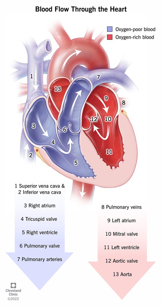
Figure 9.7 Blood flows through the heart in a series of arteries, ventricles, veins and valves.
What is the order of blood flow through the heart, step by step?
The right and left sides of your heart work together to make sure blood flows throughout your whole body. Blood flows through your heart in a series of steps. These steps take place in the space of one heartbeat — just a second or two.
On the right side
- Oxygen-poor blood from all over your body enters your right atrium through two large veins, your superior vena cava and inferior vena cava. These veins drain blood from your upper body and lower body, respectively, and directly empty it into your right atrium.
- Your tricuspid valve opens to let blood travel from your right atrium to your right ventricle.
- When your right ventricle is full it squeezes, which closes your tricuspid valve and opens your pulmonary valve.
- Blood flows through your main pulmonary artery and its branches to your lungs, where it gets oxygen and releases carbon dioxide.
On the left side
- Oxygen-rich blood travels from your lungs to your left atrium through large veins called pulmonary veins. These veins directly empty the blood into your left atrium.
- Your mitral valve opens to send blood from your left atrium to your left ventricle.
- When your left ventricle is full it squeezes, which closes your mitral valve and opens your aortic valve.
- Your heart sends blood through your aortic valve to your aorta, where it flows to the rest of your body.
How much blood does your heart pump?
Your heart pumps about 2,000 gallons of blood each day. That’s enough to fill an 8-by-10-foot swimming pool.
It beats around 100,000 times daily. In an average life span of almost 79 years, your heart beats nearly 2.9 billion times.
- “2001_Heart_Position_in_ThoraxN.jpg” by OpenStax College is licensed under CC BY 3.0 ↵
- “Position of the Heart in the Thorax” by OpenStax College is licensed under CC BY 4.0. Access for free at https://openstax.org/books/anatomy-and-physiology/pages/19-1-heart-anatomy ↵
- This work is a derivative of Anatomy and Physiology by OpenStax licensed under CC BY 4.0. Access for free at https://openstax.org/books/anatomy-and-physiology/pages/1-introduction ↵
- “Heart_diagram-en.svg.png” by ZooFari is licensed under CC BY-SA 3.0 ↵
- This work is a derivative of Anatomy and Physiology by OpenStax licensed under CC BY 4.0. Access for free at https://openstax.org/books/anatomy-and-physiology/pages/1-introduction ↵
- “2120_Major_Systemic_Artery.jpg” by OpenStax College is licensed under CC BY 3.0 ↵
- “Illu_capillary_en.jpg” by unknown author is licensed in the Public Domain. ↵
- This work is a derivative of Anatomy and Physiology by OpenStax licensed under CC BY 4.0. Access for free at https://openstax.org/books/anatomy-and-physiology/pages/1-introduction ↵
- “2131_Major_Systematic_Veins.jpg” by OpenStax College is licensed under CC BY 3.0 ↵
- This work is a derivative of Anatomy and Physiology by OpenStax licensed under CC BY 4.0. Access for free at https://openstax.org/books/anatomy-and-physiology/pages/1-introduction ↵
- CrashCourse. (2015, July 20). Blood Vessels, Part 1 - Form and function: Crash Course Anatomy & Physiology #27. [Video]. YouTube. All rights reserved. https://youtu.be/v43ej5lCeBo ↵
- “Surface Anatomy of the Heart” by OpenStax College is licensed under CC BY 4.0 ↵
