7.1 Introduction to Imaging and Diagnostics
Brandon Censon MPH, CPH, RRT-NPS, CPFT, CPT
Medical imaging and diagnostics refer to a variety of technologies used to evaluate the human body in order to diagnose, monitor, or treat medical conditions. The type of technology used depends on the area of the body being monitored or treated, as well as the specific disease, illness, or condition. It is important to note that professionals in these roles do not diagnose. Rather, they obtain important clinical information that helps the clinical care team make informed decisions about patient care.
Within medical imaging, there are several common types, including X-rays, magnetic resonance imaging (MRI), ultrasound, computed tomography (CT) scans, and nuclear medicine imaging. Each imaging type uses specific technology and modalities.
X-rays
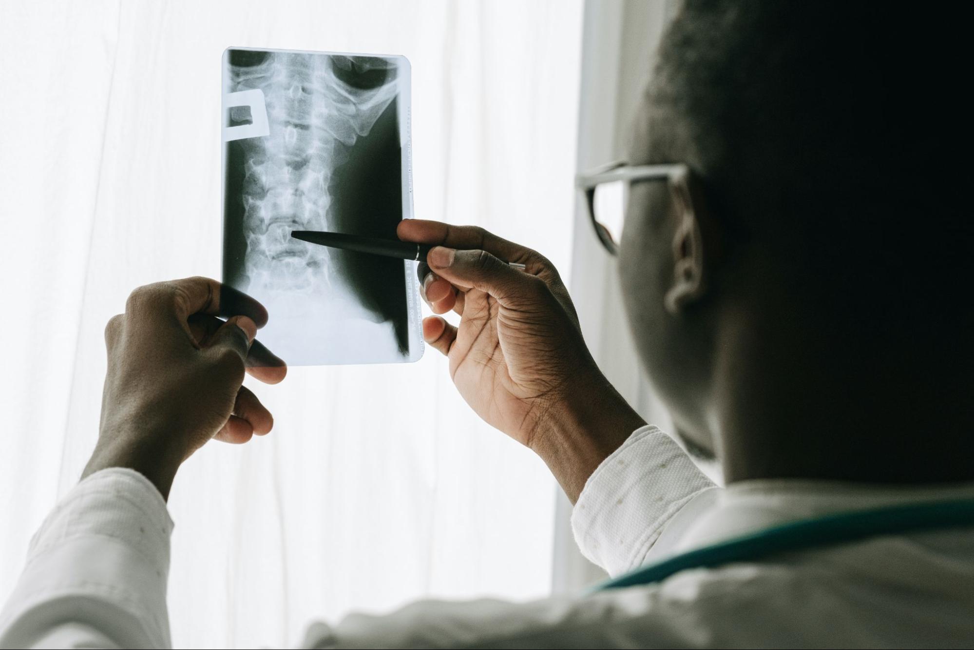
X-rays use ionizing radiation to create images of the body. This is a non-invasive and painless procedure that allows the healthcare team to diagnose diseases and monitor therapy. The images generated by X-rays help support medical and surgical treatment planning, and they can also be used during procedures to verify the correct placement of catheters or other medical devices inside the body. When the X-ray is activated, radiation travels through the body, and the amount absorbed by the tissues depends on their radiological density. For example, bones have a higher radiological density, which allows them to absorb more X-rays and produce high-contrast images. On the black background of the X-ray image, bones appear white. In contrast, tissues that are less radiologically dense, such as muscles or the air-filled lungs, appear as various shades of gray.

Computed Tomography
Computed tomography (CT) uses traditional X-ray imaging along with a computer to process a series of cross-sectional images, or slices. The images produced by CT are far more detailed than traditional radiographs, allowing the healthcare team to view structures from many different angles. These cross-sectional images can then be stacked to create a three-dimensional image of the structure. During a CT scan, the patient lies on a bed that moves in and out of a fixed X-ray tube, which rotates while capturing the images. As it rotates, X-rays are emitted and pass through the patient’s body. The X-ray detectors on the opposite side of the X-ray source pick up the radiation, and the image is transmitted to a computer for processing. Similar to traditional X-rays, radiologically denser structures appear white, while less dense structures appear in various shades of gray.
Magnetic Resonance Imaging
Magnetic resonance imaging (MRI) uses strong magnetic fields and radio waves to produce images of the internal structures of the human body. The magnetic resonance signal is generated by the protons in fat and water molecules. During the MRI exam, an electrical current passes through coiled wires, creating a temporary magnetic field in the patient’s body. Radio waves are then sent from a transmitter in the MRI machine, and the signals are used to create digital images of the scanned area. MRIs are commonly used to diagnose diseases and monitor treatments. MRIs are particularly useful for evaluating soft tissues, such as the brain, nerves, muscles, tendons, and ligaments, as these structures can be viewed more clearly than with traditional X-rays or CT scans.
Ultrasound Imaging
Ultrasound imaging is a specialized technique that uses high-frequency sound waves to view internal structures of the body. The images captured during an ultrasound are in real time, which is particularly useful for evaluating the flow of blood and other movements. Unlike traditional X-ray imaging, ultrasound does not use ionizing radiation. The images are produced by the reflection of sound waves off structures within the body.
Other benefits of ultrasound imaging include its non-invasive nature, lower cost, and ability to be used in various settings. However, there are some limitations. For instance, ultrasound waves can be disrupted by air or gas, making it difficult to evaluate structures surrounded by air. Additionally, a patient’s body composition can affect the clarity of the image, as ultrasound waves are weakened when passing through thick layers of tissue.
Ultrasound is an essential tool used by healthcare providers to diagnose, evaluate, and treat a variety of conditions. Common procedures that utilize ultrasound imaging include:
- Abdominal Ultrasound: Used to evaluate the various tissues and organs in the abdominal cavity.
- Breast Ultrasound: Helps visualize breast tissue to identify breast cancer.
- Fetal Heart Rate Monitoring: Doppler ultrasound allows for non-invasive monitoring of a fetus’s heart rate in the womb.
- Fetal Ultrasound: Used to evaluate the growth and development of a fetus throughout pregnancy.
- Ultrasound-guided Needle Placement or Intravenous Catheter Placement: Helps healthcare providers ensure that they are placing needles or intravenous catheters into specific tissues or vessels.
- Cardiac Echocardiogram: Provides images of the heart and blood flow.
Nuclear Medicine Imaging
Nuclear imaging is a type of medical imaging that uses tiny amounts of radioactive material combined with a carrier molecule. This combination is referred to as a radiotracer. Nuclear medicine imaging is a valuable tool for evaluating, diagnosing, and treating various conditions. When the radioactive material is swallowed or injected into the body, the radiotracer accumulates in the area being evaluated. This technique is often used to examine areas that are inflamed or may be suspicious for cancer. Cancerous cells typically consume large amounts of energy, especially glucose. The imaging device used in nuclear medicine detects this excess energy, creating an image that shows the location of the radiotracer.
In addition to helping with diagnoses, nuclear medicine can also be used to treat certain types of cancers and other conditions. The radioactive material attaches to specific cells and delivers a prescribed dose of radiation, which in turn destroys the cells. Nuclear medicine imaging can also be used to evaluate heart function and blood flow, especially after a heart attack, to assess any damage. It can also be used to evaluate the lungs, which is helpful when a blood clot in the pulmonary circulatory system is suspected. Other body systems that can be evaluated using nuclear medicine imaging include the gastrointestinal, endocrine, and neurological systems.
Because many diseases and conditions begin at the cellular level, nuclear medicine allows healthcare providers to detect diseases earlier in their course. With earlier detection, the healthcare team can respond more quickly, providing treatment and therapy that may help stop or slow the spread of the disease.
Attributions
- Figure 7.1: image released under the Pexels License
- Figure 7.2: image released under the Pexels License
The act or practice of diagnosis .
Identify or determine the nature and cause of a disease or a problem, typically through the examination of symptoms, medical tests, and other relevant information.
Refers to a medical examination where pictures are generated of internal structures of the body.
A medical technique that uses electromagnetic radiation to create detailed pictures of the inside of the body, aiding in the diagnosis of various health conditions.
A medical procedure or technique that does not require the insertion of instruments or devices into the body.
Also known as CT. A diagnostic imaging procedure that is used to produce images of internal structures of the body.
Abbreviated as MRI. A diagnostic imaging tool that produces 3D images of internal structures of the body.
A specialized type of imaging that uses high-frequency sound waves to view internal structures of the body.
Use of radioactive materials to visualize how organs and / or tissues are functioning. Often times this is used in the treatment and monitoring of a patient and their condition.
